Bioluminescence — the light made by fireflies, deep-sea shrimp, and glowing bacteria — is not magic or heat; it is chemistry. At its core is a two-actor reaction: an enzyme called luciferase brings together a light-producing substrate molecule called luciferin and oxygen, and the result is a new molecule, oxyluciferin, plus a photon of visible light. In fireflies, the reaction adds a third essential player, ATP, the cell's energy currency, which activates luciferin before oxygen is ever involved. That same molecular pathway, once understood, has been borrowed by scientists and engineered into two families of tools: reporter-gene imaging that lets researchers watch cancer cells and other biological processes inside living animals in real time, and biosensors that detect toxic contaminants and bacterial contamination in food and the environment.
The Chemical Spark: How Bioluminescence Works
Wherever bioluminescence appears in nature, the same basic recipe applies. NOAA's Ocean Exploration fact sheet describes it plainly: luciferase helps bond together the substrate luciferin and oxygen, and the reaction creates two products — oxyluciferin and light. The enzyme itself is a catalyst, not a fuel; it is recycled after the reaction and can be used again. That is why a firefly can flash repeatedly without exhausting its light-making machinery.
What varies enormously across species is the substrate. Different organisms use different luciferin molecules, a fact NOAA highlights as evidence that the ability to make light evolved independently in different creatures at different times. The reaction can also happen in different places: in some shrimp, it occurs outside the organism; in some animals, inside their cells; and in others, it is produced by luminous bacteria living within them. The enzyme is specific to its luciferin substrate, so a luciferase from one organism will not necessarily work with another organism's luciferin.
One striking feature of this chemistry is its efficiency. Because the energy of the reaction is released as light rather than heat, bioluminescence is often called "cold light" — a glow with minimal heat generation. That property matters greatly for the applications that follow, where a light signal must be generated inside delicate living tissue without cooking it.
Firefly Bioluminescence: A Step-by-Step Mechanism
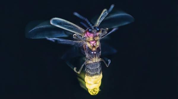
The firefly system is the best-understood example, and it unfolds in two defined chemical steps. First, luciferin reacts with ATP, and the cell's energy is spent attaching an AMP group to the substrate, forming an activated intermediate called luciferyl adenylate (also written luciferyl-AMP), while releasing pyrophosphate as a byproduct. Second, that intermediate reacts with molecular oxygen, producing oxyluciferin, AMP, and light. The glow itself comes from a quantum transition: the oxidation leaves oxyluciferin in an electronically excited state, and as the molecule relaxes back to its ground state, it releases the excess energy as a photon of visible light.
The pathway is not perfectly tidy. A side reaction diverts some of the luciferyl adenylate intermediate to react with oxygen in a different way, forming hydrogen peroxide and a compound called dehydroluciferyl-AMP instead of light. Roughly one-fifth of the intermediate is lost to this unproductive branch, which is why firefly luciferase is described as generating light from luciferin in a multistep process rather than a single clean conversion.
The Firefly's Glow: A Molecular Pathway to Light
To see how the pieces fit together, trace one luciferin molecule through the firefly reaction from chemical input to light output. Each stage has a distinct molecular actor with a distinct job.
| Stage | Molecule involved | Role at this stage |
|---|---|---|
| 1. Activation | Luciferin + ATP | Luciferin is the light-producing substrate; ATP supplies the energy to activate it, forming the intermediate luciferyl adenylate and releasing pyrophosphate. This activation is the first step of the firefly reaction. |
| 2. Catalysis | Luciferase | The enzyme catalyzes the reaction; it is specific to the luciferin substrate and is recycled afterward, ready for another cycle. |
| 3. Oxidation | Oxygen | Molecular oxygen oxidizes luciferyl adenylate — an oxygen-dependent step — yielding oxyluciferin and AMP. |
| 4. Light emission | Excited oxyluciferin | Oxyluciferin is formed in an electronically excited state; as it returns to its ground state it releases a photon of visible light, producing "cold light" with minimal heat. |
The sequence explains why each ingredient is indispensable. Remove ATP and luciferin never becomes the activated intermediate. Remove oxygen and the oxidation that creates excited oxyluciferin cannot occur. Remove luciferase and the reaction proceeds too slowly to produce a usable flash. It also explains the side branch: about 20 percent of the luciferyl adenylate is diverted by oxygen into hydrogen peroxide and dehydroluciferyl-AMP, a pathway that yields no light at all.
Seeing the Unseen: Bioluminescence in Bioimaging
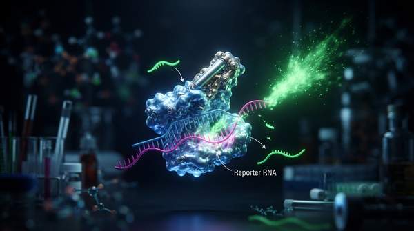
The firefly pathway has a property that makes it remarkably useful in the laboratory: light only appears when luciferase, luciferin, ATP, and oxygen are all present in a living cell. Scientists exploit this by inserting the gene for luciferase into cells or organisms as a reporter gene. Wherever and whenever that gene is expressed, the cell manufactures luciferase — and when luciferin is supplied, the cell lights itself up. The glow becomes a readable proxy for a biological event.
According to the life-sciences reagent company Biotium, this technique enables researchers to study biological processes in real time, in vivo, and has been developed over the past few decades primarily in small animals for molecular imaging. Firefly luciferase is the most commonly used bioluminescent reporter, though luciferases cloned from jellyfish, coral, beetles, and bacteria have also proved useful, and Renilla luciferase is another frequently used reporter enzyme.
A scientific review of bioluminescence systems describes bioluminescence imaging as an economical and noninvasive technology designed for real-time examination of biological processes at the molecular level, with D-luciferin serving as the substrate for the reaction catalyzed by firefly luciferase. Because the light comes from the cells themselves, researchers can track labeled cells — such as proliferating cancer cells — inside a living animal without surgery or destructive sampling, watching a tumor's behavior unfold over time in the same subject.
Guardians of Health: Bioluminescence in Environmental and Food Safety
The same chemistry also powers detection outside the body. The review of bioluminescence applications notes that bioluminescence-based sensors provide a specific, cost-effective, and reliable assessment method for evaluating contaminants, and that they have been applied to detect toxicants in food at minor levels and to monitor bacterial contamination and bacterial counts in food. In these systems, the presence of a target substance or microorganism is linked to the luciferase reaction, so the intensity of emitted light becomes a quantitative readout: more light signals more of the target.
Two features make the approach attractive for monitoring work. The method can identify toxicants at extremely low concentrations, catching contamination before it becomes visible or dangerous. And because the signal is generated chemically rather than by shining an external lamp on the sample, the review characterizes this detection as faster and more sensitive than fluorescence-based alternatives. The applications extend beyond food: the same review points to environmental monitoring and medical diagnostics as fields where harnessing the bioluminescence mechanism is opening new possibilities.
Harnessing Nature's Light: Future Directions
Bioluminescence, particularly the firefly system, serves as a robust and versatile platform for developing advanced tools in biomedical research and environmental diagnostics. The logic connecting the two is the ATP-activated, oxygen-dependent pathway traced above: a reaction that requires a living cell's own energy and chemistry is ideal both for reporting on those cells in imaging and for quantifying contaminants in sensing.
For readers deciding whether these tools matter to their work, the concrete indicator to watch is the continued development and adoption of bioluminescence-based reporter systems in research labs and commercial biosensing products — from firefly-luciferase reporter assays tracking tumor cells in small animals to sensors detecting food toxicants at minor levels and counting bacterial contamination. Each new reporter construct or commercial biosensor built on the luciferin–luciferase reaction is a measurable sign that nature's cold light is still being put to work.